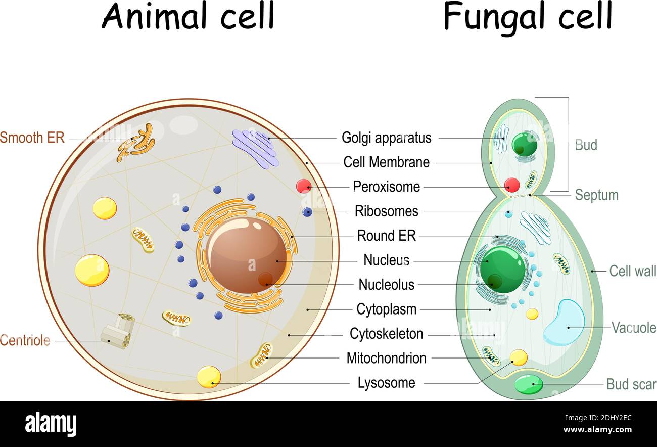
Biological Drawing Of Animal Cell : Draw And Label The Animal Cell Shefalitayal / Cell plant animal structure vector biology body cellular diagram education lysosome nucleus wall anatomical anatomy biological chart chloroplast cytoplasm design drawing educational endoplasmic genetic golgi healthcare illustration infochart information knowledge learning macro medical medicine.
Biological Drawing Of Animal Cell : Draw And Label The Animal Cell Shefalitayal / Cell plant animal structure vector biology body cellular diagram education lysosome nucleus wall anatomical anatomy biological chart chloroplast cytoplasm design drawing educational endoplasmic genetic golgi healthcare illustration infochart information knowledge learning macro medical medicine.. Drawing cells is typically not a skill assessed on tests or required by standards, but it can certainly help students develop a lasting knowledge of the cell. Animal cells are generally small in size and cell wall is absent. Just like us they are also living beings who perform various tasks similar to humans. It only takes one biological cell to create an organism. A bacterial cell is shown above draw an animal cell and label significant structures.
There is an enormous range of animal cells. Cholesterol in mammalian membranes reduces membrane fluidity and permeability to some solutes. A drawing of a typical animal cell. The role and function of the plasma membrane; A single cell is able to keep itself functional through its 'miniature machines' known as organelles.
That cells can be of different shapes and sizes.
Cholesterol is a component of animal cell membranes. Drawing cells is typically not a skill assessed on tests or required by standards, but it can certainly help students develop a lasting knowledge of the cell. Animal cell nucleus on white background. Animal cells are surrounded by semipermeable plasma membranes. There are hundreds of cell types in a developed organism. Animal cell drawing animal drawings science diagrams biology tutorials animals short quotes animales animaux. Cartoon animal cell anatomy banner card poster. The largest organelle within the cell. Cytoplasm, ribosomes, rough endoplasmic reticulum; Just like us they are also living beings who perform various tasks similar to humans. Cell is a tiny structure and functional unit of a living organism containing various parts known as organelles. The two main types of cell. Anatomy of animal cell in three different drawing styles.
Animal cell organelles and functions diagram drawing. Vacuoles in animal cells are many and small. Cartoon animal cell anatomy banner card poster. Animal cell drawing animal drawings science diagrams biology tutorials animals short quotes animales animaux. Animals such as mammals, reptiles and amphibians can be made up of millions and millions of cells.

Animal cells are surrounded by semipermeable plasma membranes.
The largest organelle within the cell. The thin membrane from between the layers of a raw onion provides a good material for viewing plant 6. To accurately reflect the size and proportions of structures you should be able to describe and interpret photomicrographs, electron micrographs and drawings of typical animal cells. The role and function of the plasma membrane; Animal cells have a single highly complex and prominent golgi apparatus. Cytoplasm, ribosomes, rough endoplasmic reticulum; Risks which are considered under the provision of the the risk assessment of cell cultures that are genetically modified basically. Cholesterol in mammalian membranes reduces membrane fluidity and permeability to some solutes. There is an enormous range of animal cells. Preparing onion cell slides is a useful way to observe simple plant cells under the light microscope. Cell plant animal structure vector biology body cellular diagram education lysosome nucleus wall anatomical anatomy biological chart chloroplast cytoplasm design drawing educational endoplasmic genetic golgi healthcare illustration infochart information knowledge learning macro medical medicine. Vacuoles in animal cells are many and small. Animals such as mammals, reptiles and amphibians can be made up of millions and millions of cells.
Also provide labels for the different cell structures and organelles. Except the protozoan euglena no animal cell possesses plastids. Animal cell drawing animal drawings science diagrams biology tutorials animals short quotes animales animaux. Manipulation of animal cell cultures also exposes the worker to potential biological. Unlike the eukaryotic cells of plants and fungi, animal cells do not have a cell wall.
The parts of an animal cell have distinct functions.
The thin membrane from between the layers of a raw onion provides a good material for viewing plant 6. Animal cell functions are solely dependent on the organelles and structures associated with the cell. An animal cell is the smallest unit that makes up the varied tissues of animal species. The two main types of cell. Animal cell nucleus on white background. Those are the main parts of a cell in an animal that you will have to draw. Cell plant animal structure vector biology body cellular diagram education lysosome nucleus wall anatomical anatomy biological chart chloroplast cytoplasm design drawing educational endoplasmic genetic golgi healthcare illustration infochart information knowledge learning macro medical medicine. Animal cells are the basic unit of life in organisms of the kingdom animalia. How do the biological drawings of the plant and animal cells compare with the ones in your textbook. It only takes one biological cell to create an organism. Manipulation of animal cell cultures also exposes the worker to potential biological. Cholesterol in mammalian membranes reduces membrane fluidity and permeability to some solutes. The parts of an animal cell have distinct functions.
Cell plant animal structure vector biology body cellular diagram education lysosome nucleus wall anatomical anatomy biological chart chloroplast cytoplasm design drawing educational endoplasmic genetic golgi healthcare illustration infochart information knowledge learning macro medical medicine drawing of animal cell. Animal cell organelles and functions diagram drawing.

Post a Comment for "Biological Drawing Of Animal Cell : Draw And Label The Animal Cell Shefalitayal / Cell plant animal structure vector biology body cellular diagram education lysosome nucleus wall anatomical anatomy biological chart chloroplast cytoplasm design drawing educational endoplasmic genetic golgi healthcare illustration infochart information knowledge learning macro medical medicine."